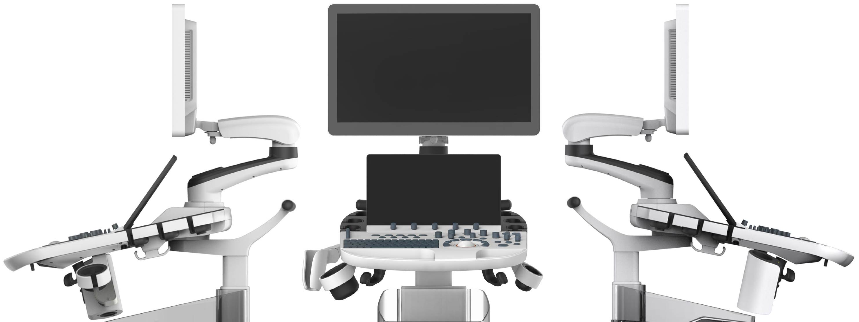
P50
Define Your Vision
Define Your Vision
Easily accomplish more with SonoScape’s new P50 ultrasound system. Incorporating single crystal clarity, automatic corrections and calculation, and user defined flexibility promises a confident diagnostic experience as well as opening new doors of opportunity for ultrasound use.

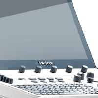
Taking into consideration the evolving expectations and needs for ultrasound, the P50 is a slim and unobtrusive trolley system that is comfortable in tight, congested spaces with little room to work in. Providing everything you need for a comfortable examination in a small space for both you and your patient.
High Sensitivity Touch Screen
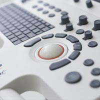
Simplified Control Panel
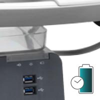
Power Assistant Battery Operation
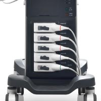
All Active Transducer Sockets
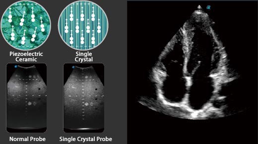
Wide band single crystal probes greatly improve signal ratio, acquire stunning images and provide superior sensitivity and resolution for both the near and far fields.
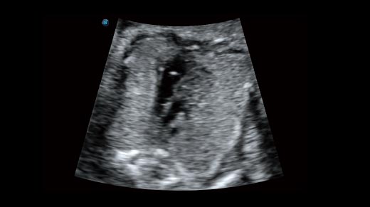
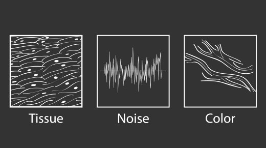
The new generation μ-Scan imaging technologies give you better image quality by reducing noise, improving signal strength and improving visualization.
Dynamic color improves upon already existing color Doppler technologies for a clear capture of color flow and detail visualization of even tiny veins with lower velocities.

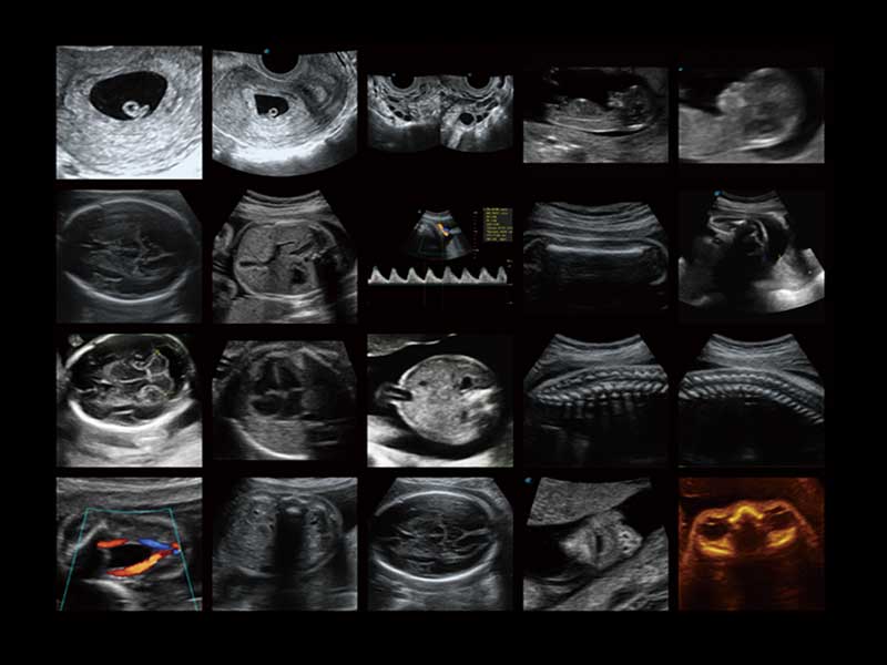
P50, is leading-edge ultrasound system that can meet the demands of any clinical setting. You can experience a superior performance in multi-dimensional imaging for a full range clinical applications – abdominal, breast and cardiovascular.
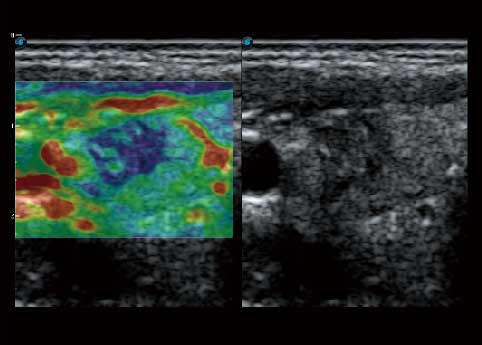
By understanding that tissue stiffness varies depending on the type of tissue, we can use C-xlasto Imaging to easily find abnormalities and tumors within soft tissue. The differences in tissue responses are detected and visualized in real-time by the elastography algorithms through different representations, which can be particularly helpful in analyzing breast, thyroid and musculoskeletal structures.Predominately used only in linear probes, SonoScape’s new transvaginal and bi-plane probe for gynecology and urology are breaking the mold and expanding elastography applications.
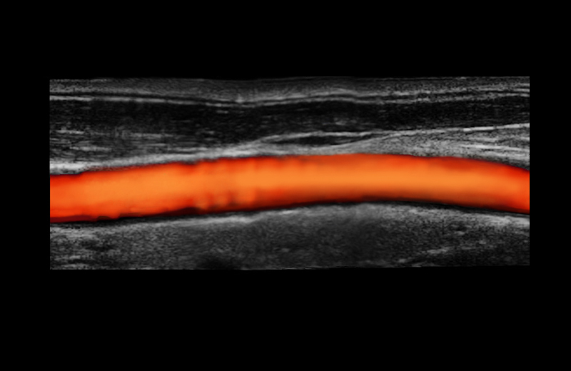
With the combination of color flow and real-time panoramic, visualizing the blood flow of an entire vein or artery is now an easy task. Accomplished in real-time for the convenience of the sonographers, any mistakes can also be easily back tracked and corrected without interrupting the scan.
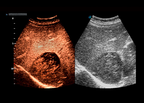
Contrast Imaging on P50 makes full use of the infra harmonic signal and second harmonic signal to improve the image resolution and deep penetration. What’s more, the Dynamic Acoustic Control technology effectively controls the acoustic pressure for the contrast agent, decreasing the required agent dose and assures uniform image quality, guaranteeing longer contrast agent duration and better lesion perfusion of delayed phase observation.

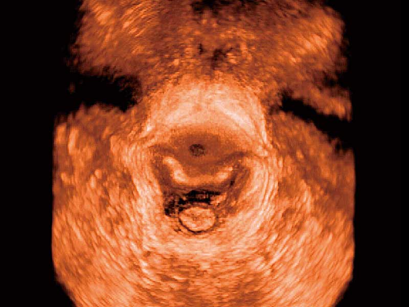
P50 has superior image quality, automated measurement tools, and a variety of volume technologies to provide ideal solutions for clinical examinations such as pregnancy examinations, and gynecologic disease diagnosis. With a new 4D transvaginal probe, P50 helps you to see and detect fetal abnormalities, and significantly improves your diagnostic confidence during your examinations.

A unique transparent 3D anatomical image of the fetus for improved initial anatomical review. By using this new application, the system can create completely different fetal images from conventional ultrasound images, which can depict the fetal's intracorporeal anatomical structure.
Working in conjunction with SonoScape’s latest transvaginal probes, trans-perineal 4D pelvic floor ultrasound provides useful clinical assessment of the impact of vaginal delivery on the female anterior compartment. Allowing doctors to judge whether the pelvic organs prolapsed or not, the extent of porlapse, and determining whether the pelvic muscles tore correctly.
S-Guide gives the user an extensive list of example obstetric ultrasound images as reference guides and a convenient check list system to keep track of their progress during their obstetrics examination.
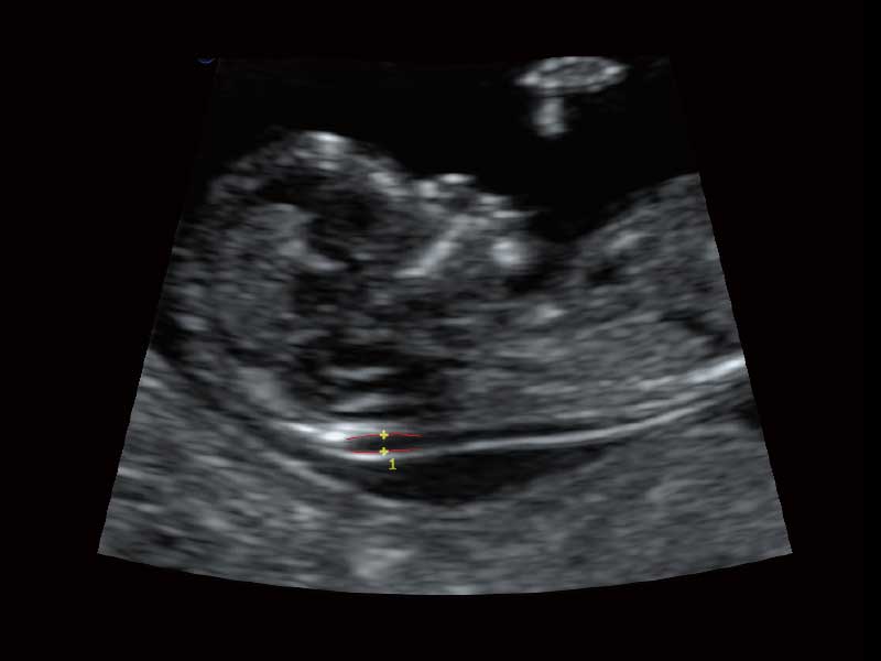
Auto NT automatically traces and then calculates the thickness of the nuchal posterior transplant layer for a faster evaluation.
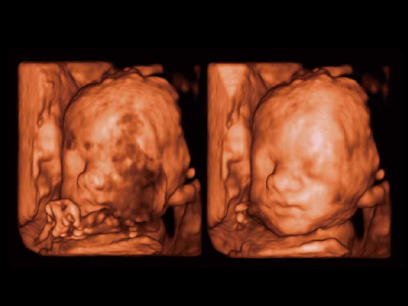
Automatically removes masking layers in front of the fetus’s face for a clearer visual of the fetus’s face.
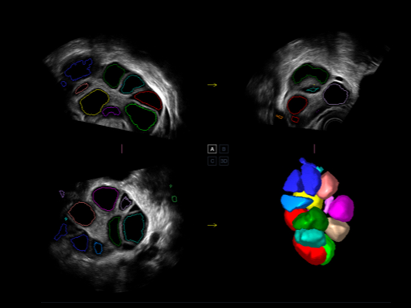
AVC Follicle automatically identifies how many follicles are present and calculates their individual volumes.

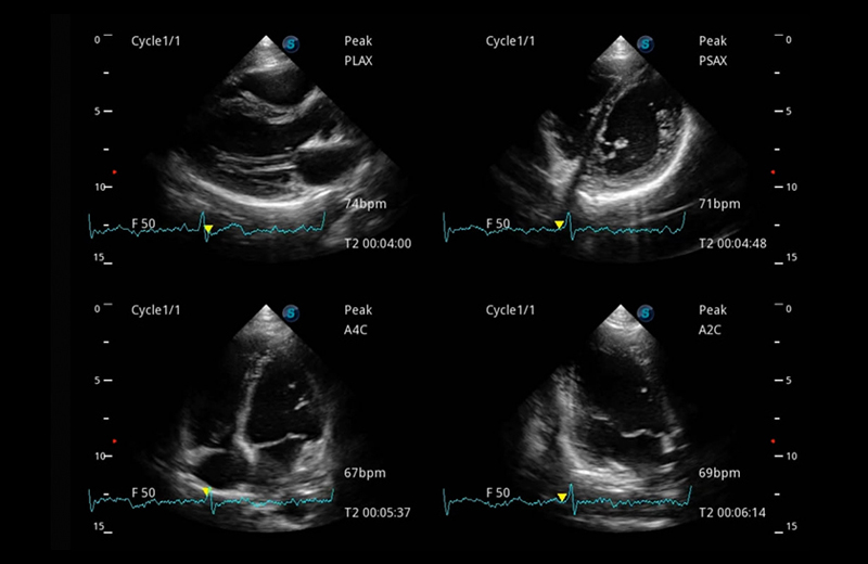
P50 provides clear 2D clinical images and Doppler sensitivity to assess critical cardiac performance. Compatible with SonoScape’s single crystal probes, the P50 can provide images with better resolution and penetration in Cardiac diagnosis.
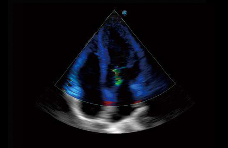
Tissue Doppler Imaging allows clinical doctors to quantitatively evaluate local myocardial movements and functions, facilitating them with the ability to analyze and compare the motions of the different parts of the patient’s heart.
Stress echocardiography is the combination of 2D echocardiography with a physical, pharmacological or electrical stress of the patient. It also then provides users with report management tools such as configurable template editor, multiple loops to select one for storage, wall motion scoring, stress echo report, etc.
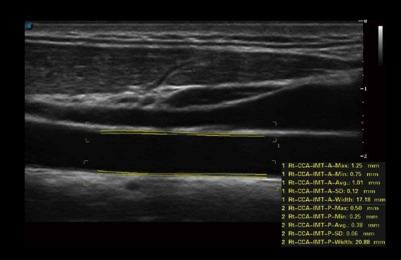
Auto IMT is used when determining the level of vascular sclerosis present in the patient by automatically tracing and calculating the thickness of the carotid vessels. What distinguishes the P50 is that it provides an instant and accurate Mean and Max index at the touch of a single button.
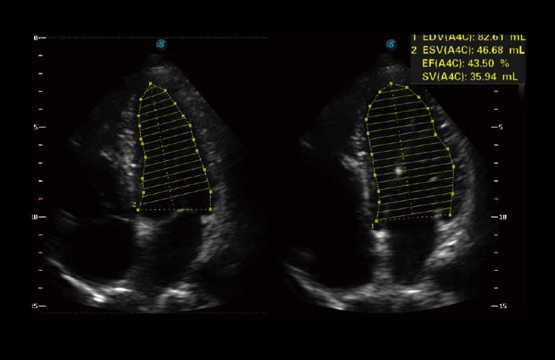
Automated 2D Cardiac Quantification is a fully intelligent trace function for endocardium with 19 easily-adjustable points providing rapid access to proven 2D EF and volumes.

Breast

C-xlasto Imaging

Cardiac

Cardiac

Carotid Artery

TDI

Fetus Cardiac

Fetus Spine

Pelvic Floor

Uterus

Gallblader

Real-time Panoramic

Spermatic Vein

Thyroid
SonoScape Medical Corp. stands as a prominent innovator in medical technology, specializing in ultrasound medical imaging, endoscopic diagnosis and treatment, minimally invasive surgery (MIS), and cardiovascular intervention solutions. Offering professional medical solutions and support in over 170 countries, SonoScape is driven by a passion for continuous innovation, unlocking life's potential and paving the way for boundless advancements in healthcare.
Copyright © SonoScape Medical Corp. All Rights Reserved. 粤ICP备20054866