Marvo S80 Elite
Ultrasound System
From Boundless Exploration to Infinite Horizons
From Boundless Exploration to Infinite Horizons

Embark on a healthcare revolution with the SonoScape Marvo S80 Elite ultrasound system, merging exceptional imaging capabilities with innovative AI-driven diagnostic functions. This elite system boasts unparalleled image clarity, vivid flow hemodynamic analysis in diagnosis. Its integrated smart AI tools accelerates the process of diagnosis, ensuring rapid precise evaluations across all applications. Ergonomically refined, the Marvo S80 Elite promises supreme user engagement, facilitated by intuitive interfaces and smart automations that elevate workflow productivity, simplifying day-to-day operations, caters to diverse medical disciplines.
The C-Field+™ Architecture represents a major advancement in imaging technology. It integrates the cutting-edge C-Field Beam, innovative RF-data processing engine and the intelligent hashrate platform, allowing for brilliant image clarity and rapid signal processing. Coupled with the AI-driven Omni-Intelligence system for optimized signal selection and adaptable workflow management, it ensures superior computational capabilities and accelerated diagnostic efficiency.

Frame Rate and Spatial Resolution
Contrast Resolution
Signal-to-Noise Ratio

AI Improves Efficiency
XPUs Accelerate Computing
RF Data Boosts Processing
Marvo S80 Elite offers an aesthetic design with excellent visual effect and long-lasting performance, ensuring optimal comfort during examinations.


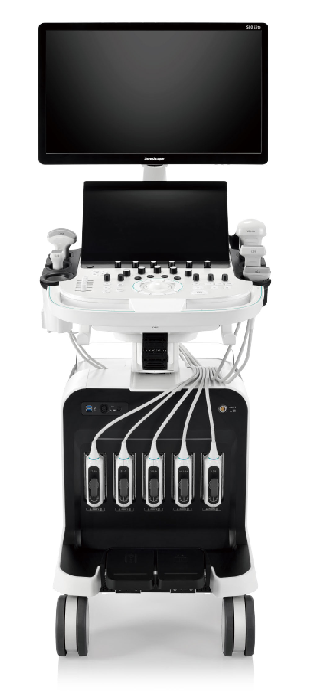
With intelligent motion boost sensor, new generation transducers support auto pick-up activation. Users can enjoy the convenience of multi-adjustable sensitivity levels tailored to their preferences, further enhancing examination flexibility and efficiency.
Automatic workflow memory helps you review the entire scanning process by automatically recording it. This feature allows to create personalized workflow steps based on the recorded data, tailored to facilitate scanning and meet quality control of workflow requirements.
Marvo S80 Elite is fully integrated into every stage of the patient management process, providing a wide range of application solutions covering from screening, diagnosis, pre-treatment, to post-treatment.
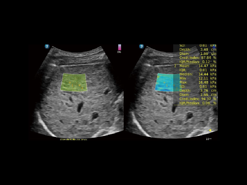
SWE (Shear Wave Elastography) empowers quantitative assessment for liver fibrosis and displays a quality map to enhance clinical diagnostic confidence.
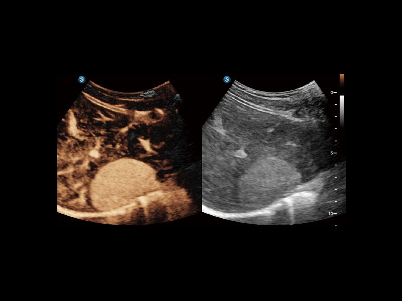
High Frame Rate CEUS dynamically displays the perfusion process of lesions in details. The combination of MFI, MFI Time and MFI Mix allows doctors to view the lesion perfusion from different perspectives and hence diagnose more easily and precisely.
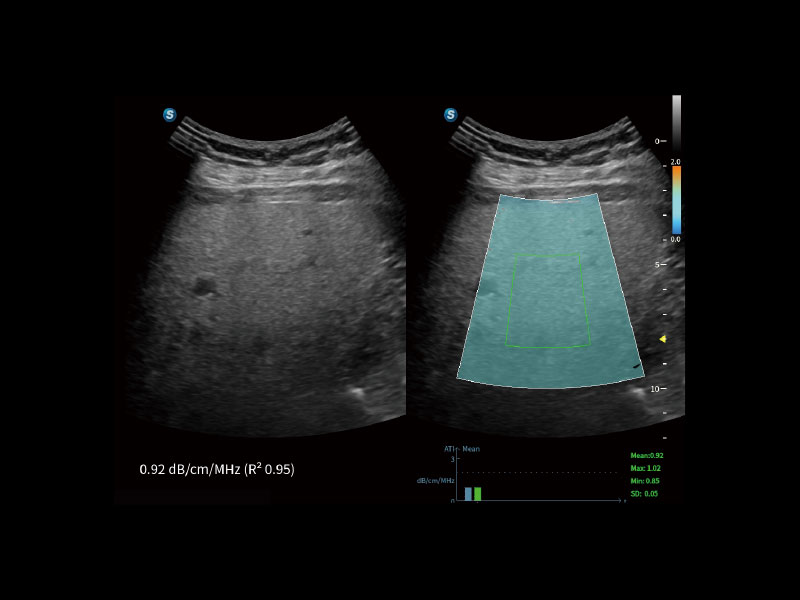
ATI (Acoustic Attenuation Imaging) enables quantitative assessment of liver steatosis through measuring the attenuation coefficient and assists clinicians in assessing the degree of steatosis for a reliable prognosis.
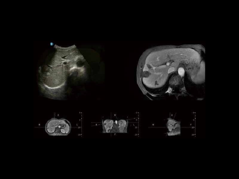
With a built-in magnetic sensor in the probe, SonoFusion eliminates the need for an external magnetic sensor, and fuses the ultrasound data with CT/MR data to achieve simultaneous display by image registration. This integration facilitates comprehensive disease analysis by clinicians.
One-stop solution for automatic standard plane acquisition and measurement. With just one click, 29 standard sections of fetal ultrasound images are intelligently identified and 13 biometrics measurements are automatically obtained with high intelligence, accuracy and efficiency, aiming for an unprecedented ease during operation.
S-Thyroid is an advanced tool in detecting and classifying suspicious thyroid lesions based on ACR TI-RADS guidelines, automatically defining the lesion boundaries and generating a report afterwards.
S-MSK enables the automatic identification of musculoskeletal joint standard sections. With a simple one-click, the desired standard planes are acquired immediately from the cine loop, and the anatomical structures are highlighted and annotated in the image.
SonoScape Medical Corp. stands as a prominent innovator in medical technology, specializing in ultrasound medical imaging, endoscopic diagnosis and treatment, minimally invasive surgery (MIS), and cardiovascular intervention solutions. Offering professional medical solutions and support in over 170 countries, SonoScape is driven by a passion for continuous innovation, unlocking life's potential and paving the way for boundless advancements in healthcare.
+86-755-26722890
Room 201 & 202, 12th Building, Shenzhen Software Park Phase II, 1 Keji Middle 2nd Road, Yuehai Subdistrict, Nanshan District, ShenZhen, 518057 Guangdong, P.R. China
Copyright © SonoScape Medical Corp. All Rights Reserved. 粤ICP备20054866