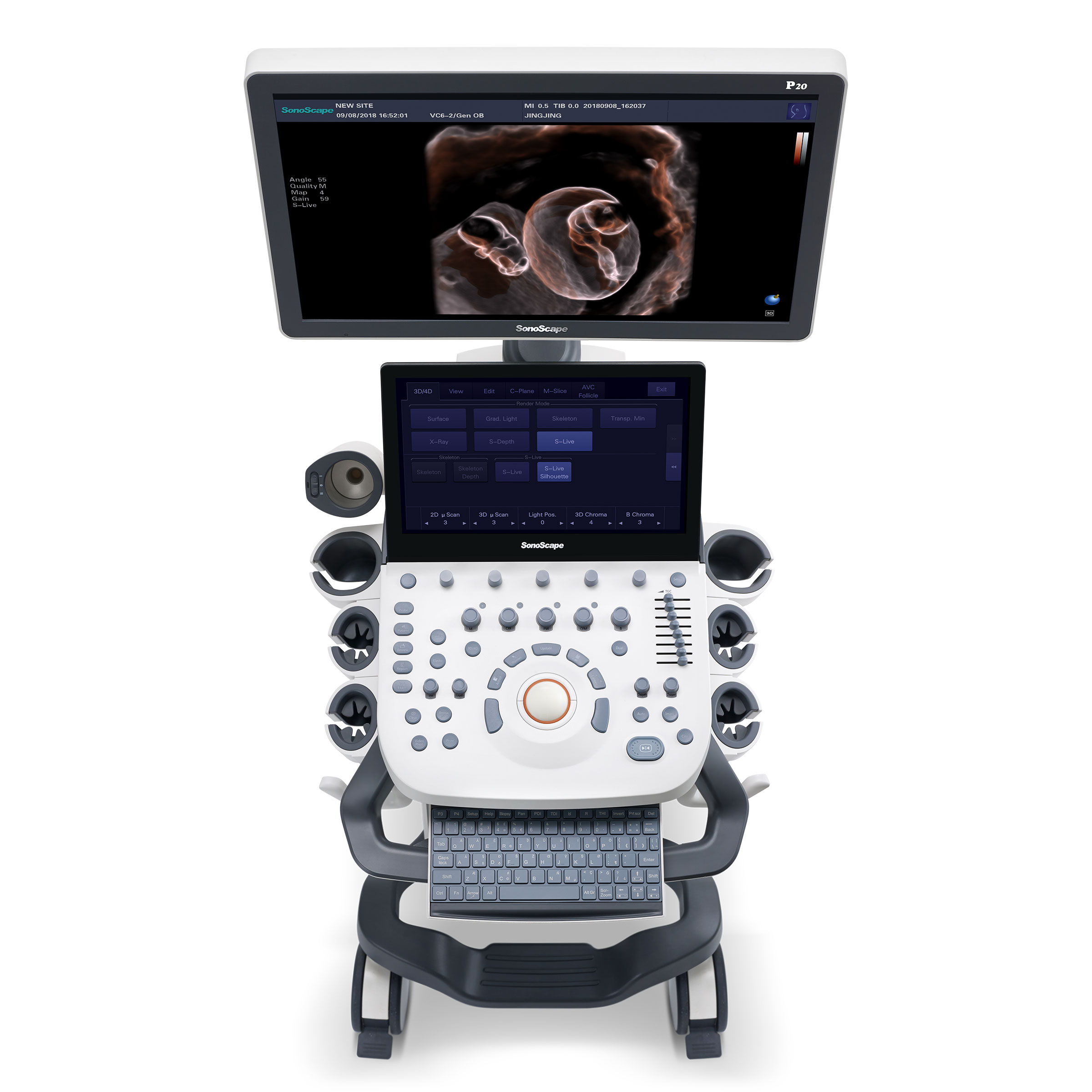
Incorporating innovative technologies, P20’s user-friendly designed with a simple operation panel, intuitive user interface and a variety of intelligent auxiliary scanning tools, will significantly improve your daily examination experience. Besides general imaging applications, P20 has entitled with diagnostic 4D technology which has an extraordinary performance in obstetrics and gynecology applications.
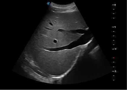
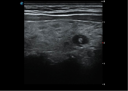
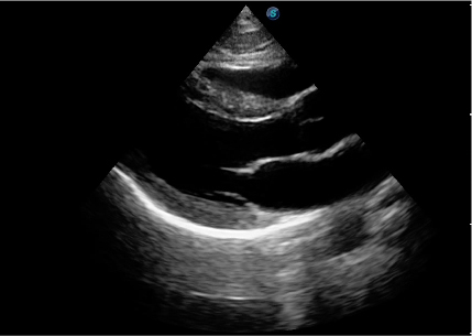
Upgraded Images with More Clarity
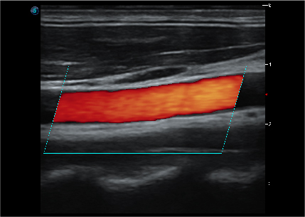
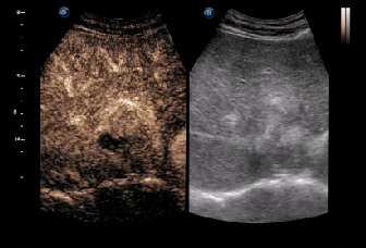
SonoScape never stop making progress in improving the image quality of its ultrasound products to enhance the confidence of diagnosis for doctors. With extraordinary images provided by P20, the anatomy structures are clearer than ever.
 |
 |
 |
 |
 |
 |
Value with Compromise, Treat with Confidence
 C-Xlasto Imaging:
With C-xlasto Imaging, P20 enables comprehensive quantitative elastic analysis. Meanwhile, C-xlasto on P20 is supported by linear, convex and transvaginal probes, to ensure good reproducibility and highly consistent quantitative elastic results.
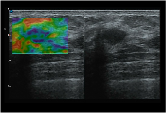
 Contrast Imaging:
Contrast Imaging with 8 TIC curves allows doctors to assess perfusion dynamics in a wide range of clinical settings, including both the location and evaluation of lesion parts.
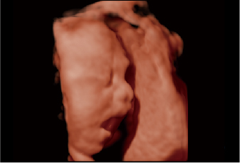 S-Live:
S-Live allows for detailed visualization of subtle anatomical features, thereby enabling intuitive diagnosis with real-time 3D images and enriching patient communication.
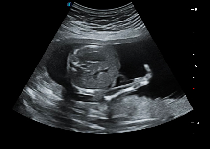
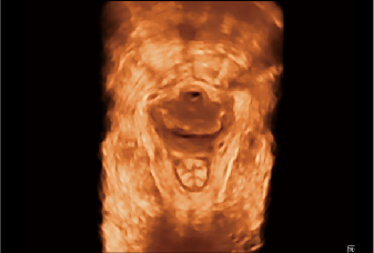 Pelvic Floor 4D:
Transperineal 4D pelvic floor ultrasound can provide useful clinical values in assessing the vaginal delivery impact on the female anterior compartment, judging whether the pelvic organs are prolapsed or not and the extent, determining if the pelvic muscles were torn accurately.
 Anatomic M Mode:
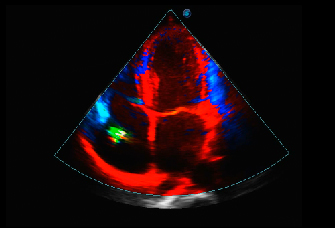
Anatomic M Mode helps you observe the myocardial motion at different phases by freely placing sample lines. It accurately measures the myocardial thickness and the heart size of even difficult patients and supports the myocardial function and LV wall-motion assessment.
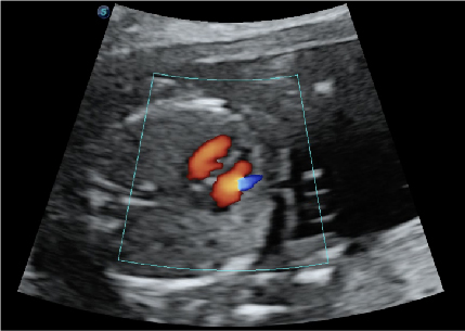
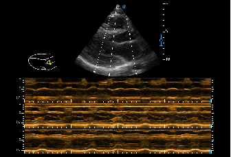
 Tissue Doppler Imaging:
P20 is endowed with Tissue Doppler Imaging which provides velocities and other clinical information on myocardial functions, facilitating clinical doctors with the ability to analyze and compare the motions of different parts of the patient's heart.
Easily Accomplish More with One Click
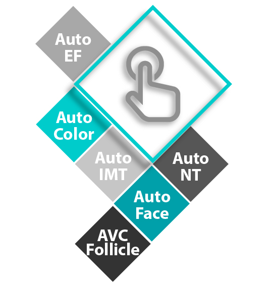
SonoScape's automatic tools can help you to achieve more. Sonographers can access the result with only ONE click. The automatic tools includes Auto NT, Auto Face and AVC Folliclein OB/GYN application, also Auto EF, Auto IMT in Cardiovascular.