The Full Shared Service System for Veterinarian's Imaging Need
Veterinary Color Doppler Ultrasound System
Veterinary Color Doppler Ultrasound System
All creatures are great and small. Animals are the closest friends and most trusted partners of human beings. SonoScape has always been devoted to exploring the dedicated diagnostic imaging solutions for veterinary needs. Starting a new phase in ultrasound imaging, SonoScape rolls out the brand new ProPet series systems to provide professional veterinary solutions catering to a wide variety of species ranging from small pets to large animals. ProPet 70 is a trustworthy ultrasound solution of veterinarians aiding in the acquisition of decisive clinical evidence and informative evaluation across a wide range of animals types with intuitive operation. It combines the latest intelligence veterinary software and single crystal probe technology to deliver exceptional images, defining the subtlest pathology, from great danes to tiny exotics and significantly delivers better clinical outcomes with incrediable values beyond your expectations.
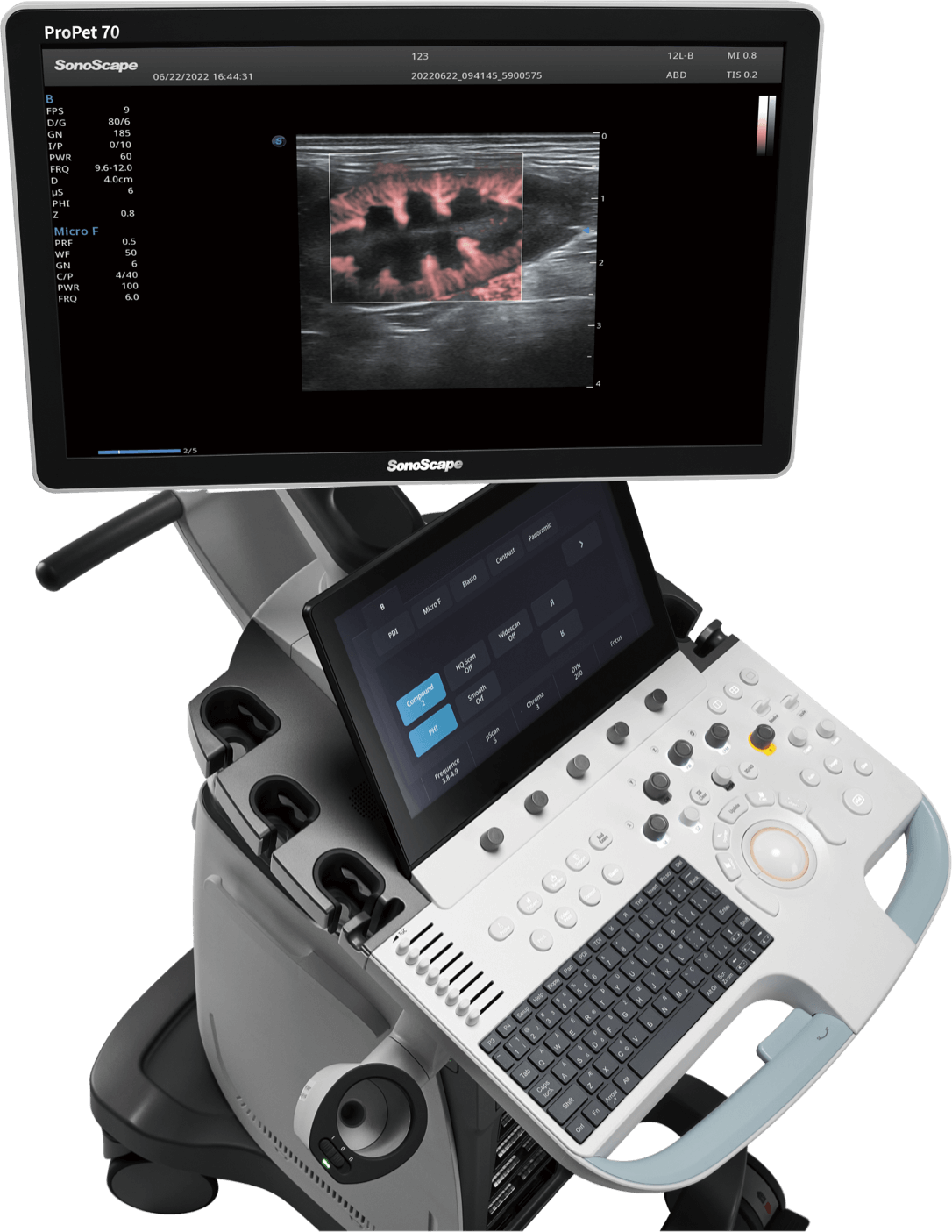
The ProPet 70 features a 23.8" full HD LED display for optional, delivering excellent contrast resolution, image clarity and vibrant color in any lighting condition
13.3" anti-glare and anti-fingerprints touch screen with 15 degree rotation
Water-proof and free of animal hairs
Power management with battery supporting 2 hours continuous scanning per charge in case of power failure
To save clinicians valuable time and energy by relieving the trouble of changing transducers frequently
The height-adjustable control panel features a simplified console design, providing intuitive interaction for a variety of veterinary examinations
With a wide range of motion, the fully articulating monitor arm adapts to your changing needs
Feline, small to medium sized canine and exotic small animals
Enhanced needle visualization technology reveals needle location within animal anatomy with no distortion when performing interventions like nerve blocks and tissue biopsies.
Precise left ventricular wall motion detection with globally 2D speckle patterns tracking provides accurate quantitative analysis including strain, strain rate, displacement, velocity, etc. on myocardial walls.
Distinguishes minute flow from overlaying tissue movement effectively, and depict hemodynamic with higher sensitivity and spatial resolution.
A straightforward template for clinicians to take multiple dynamic images at rest and after stress and make side by side comparison. Professional wall motion bulls-eye scoring and reporting is provided for further effective evaluation of animal cardiac muscle viability.
Offers a real-time tissue stiffness assessment displayed as a color map to detect potential abnormalities within normal tissues.
Automatic ejection fraction calculation based on left ventricular wall tracing and Simpson's rule saves time and efforts compared with manual measurement.
The non-linear contrast enhanced ultrasound imaging makes full use of harmonic and fundamental signals to give a more enhanced image of difficult-to-view blood flow. Provides a color coded parametric view, indicating the uptake time of contrast agents in different perfusion phases to better differentiate tissues.
Is delicately engineered to reduce speckles while improve image uniformity and enhance border continuity, providing authentic presentation of details and enhanced lesion display.
Big-sized canine, equine, farm animals and exotic big animals
Utilizes several lines of sight for optimal contrast resolution, speckle reduction and border detection, with which is ideal for superficial imaging with better clarity and improved continuity of structures.
Real-time 2D and Color/Power Doppler Panoramic Imaging enables to extend the field of view and any mistakes can also be easily back tracked and corrected without interrupting the scan.
Enables a dynamic and vivid Doppler display with high sensitivity while ensuring a realistic evidence for detections of slow flows.
3D-like color Doppler flow strengthens boundary definition of vessel walls, helping clinicians more intuitively visualize blood flow.
A newly-developed 2D improvement technology to enhance the contrast and whole image structure with better details.
One key bladder wall tracing and volume measurement from Auto Bladder can efficiently provide more accurate contour and results, which is not subject to the bladder shape and size.
Collects data with up to three sampling lines at one time to implement detailed assessment on wall motion. It greatly improves the reproducibility and accuracy of left ventricular measurement.
Uses myocardial Doppler frequency shifts to quantify myocardial tissue motion, with red and blue representing the different direction of wall movement.
The ProPet is designed for veterinarians to guarantee a very high level of professionalism in all vet applications. With the vet-dedicated software they can get an intuitive operation and accurate diagnosis in the fastest possible way.
Makes it easy to choose from a list of animal types, transducers as well as presets combination in one click.
Is designed specifically for veterinary environment with fields for animal’s name, species, breed and castration status.
Veterinaries is able to user-define animal type and corresponding graphic exam icon in system setting for specific veterinary application.
Covers a full range of applications like reproduction, cardiac and MSK, etc, providing necessary tools to the user’s needs, including Cornell & BSA animal specific formulas.
Measure
Report
Covers 200+ veterinary bodymarks and 120+ veterinary annotations including small animals, equine, farm animals, lab animals and exotic animals with more than 20 species.
Veterinary Bodymarks
Veterinary Annotations
Species
Veterinarians’ work can be burdensome due to the daily ins and outs. SonoScape strives to prepare veterinarians as much as possible so they can focus on what they are passionate about. Better user experience always was, and always will be, our goal.
Provides a fast and convenient ultrasound image transmission between ultrasound machine and smart devices.
Guides clinicians through the entire exam and provides customizable abdomen and cardiac scanning templates helping streamline workflow while increasing standardization.
Makes it possible to connect ultrasound with smart devices, laptop computer or even another ultrasound system in a remote distance and perform remote medical consultation and tutorial.
Provides intuitive one touch 2D, Color and Dopple image optimization through intelligent real-time algorithms.
It can trace the PW/CW spectrum automatically, which help doctors make measurement easily and conveniently.
ProPet 70 combines the latest intelligence software and single crystal probe technology to deliver exceptional images, defining the subtlest pathology, from great danes to tiny exotics and significantly delivers better clinical outcomes.
Canine Renal Blood Flow
Feline Gallbladder
PW Mode of Feline Heart
Canine Cardiac Blood Flow
Canine Iliac Blood Flow
Canine A4C
Aortic Arch of Canine Fetus
Canine Liver
MQA of Canine A4C
SonoScape Medical Corp. stands as a prominent innovator in medical technology, specializing in ultrasound medical imaging, endoscopic diagnosis and treatment, minimally invasive surgery (MIS), and cardiovascular intervention solutions. Offering professional medical solutions and support in over 170 countries, SonoScape is driven by a passion for continuous innovation, unlocking life's potential and paving the way for boundless advancements in healthcare.
Copyright © SonoScape Medical Corp. All Rights Reserved. 粤ICP备20054866