-
Evolved Architecture Sparks Profound Vision
-
Lucid lmaging Boosted by All-rounded Renovation
-
Intelligent Solution at Your Fingertips
-
Talented Features Inspire More Applications
-
Easeful Experience within Easy Reach
-
![]()
4times
Data Processing Competence
-
![]()
10times
Response Rate
-
![]()
2times
System Frame Rate


Lucid Imaging Boosted by All-rounded Renovation
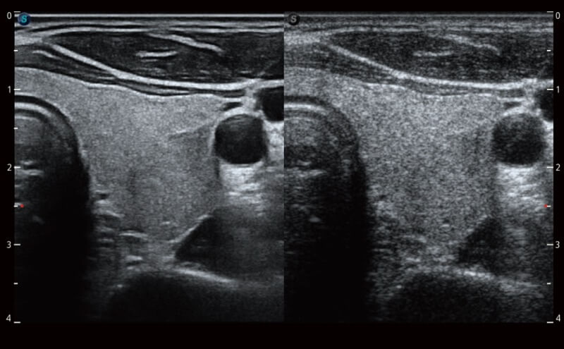
Image quality always lies at the core of definitive clinical outcomes. ELITE delivers a high-performance and lucid imaging rendered by a powerful architecture, state-of-the-art transducers, and sophisticated processing algorithms, for the next level of clarity and confidence.
μ-Scan+
A new generation μ-Scan+, available for both B and 3D/4D modes, is more delicately engineered to distinguish tissue and artifacts. In the meantime of reducing speckles, it can improve image uniformity and enhance border continuity to provide authentic presentation of details and enhanced lesion display.
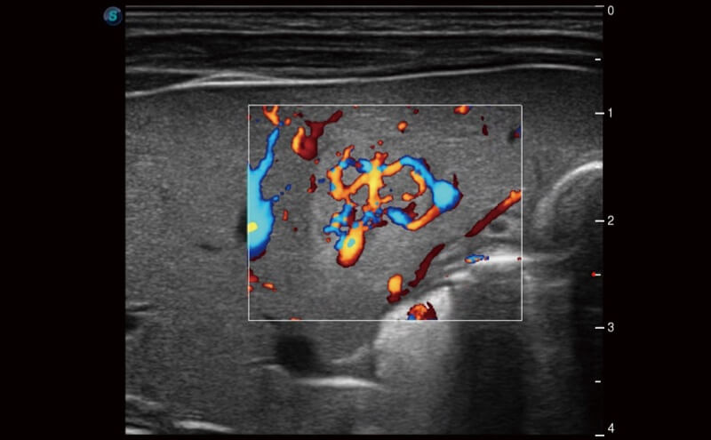
SR-Flow
The separation of blood flow and tissue signal becomes more easily with SR-Flow given the use of a highly effective filter technology. It enables a dynamic and vivid Doppler display with high sensitivity while ensuring a realistic evidence for detection of slow flows.

Micro F
Micro F provides an innovative method to expand the range of visible flow in ultrasound, especially for visualizing hemodynamic for tiny vessels. Detailed views of blood flow in relation to nearby tissue also render more diagnostic confidence to evaluate lesions and tumors.
Intelligent Solution at Your Fingertips
Routine over-repetitive exams and complicated operation are stressing out ultrasound clinicians. Intelligent solution provided by ELITE streamlines parts of the workflow to improve remarkably efficiency, with AI-powered tools including measurement, parameter adjustments, image optimization, etc.
-
![]()
S-Fetus
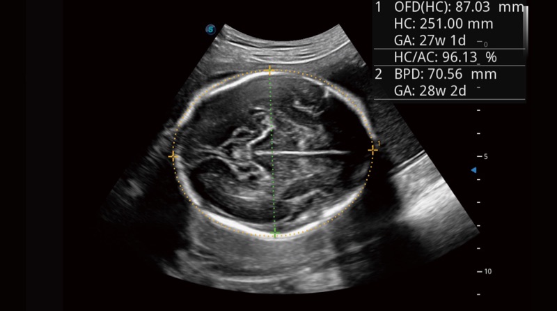
Based on a big data dependable deep learning algorithm, S-Fetus is a brilliant one-stop solution for automatic standard plane acquisition and measurement. With just one click, common fetal biometry results are obtained with high intelligence, accuracy and efficiency, aiming for an unprecedented ease during operation.
-
![]()
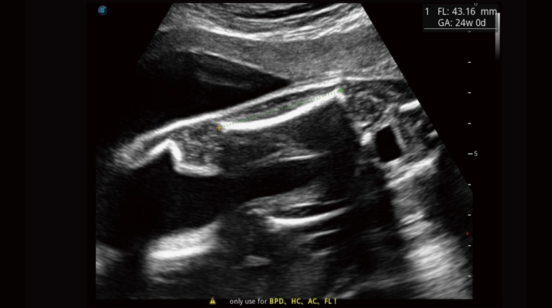
Auto OB Plus
Fast and highly efficient fetal biometry is achieved by the help of Auto OB. Meanwhile, more consistent results given by this deep learning based method can effectively reduce user-dependent variability.
-
![]()
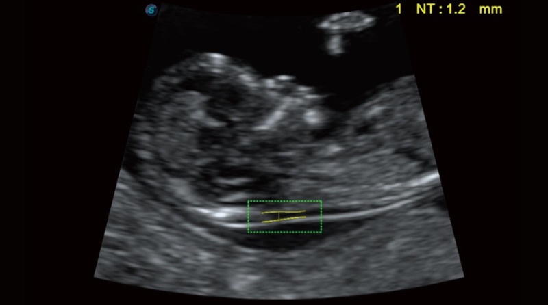
Auto NT
Auto NT provides semi-automatic, standardized measurements of the nuchal translucency thickness in 2D image and reduces operator dependency on the results.
-
![]()
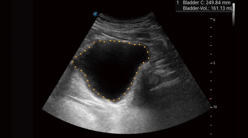
Auto Bladder
One key bladder wall tracing and volume measurement from Auto Bladder can efficiently provide more accurate contour and results, which is not subject to the bladder shape and size.
-
![]()
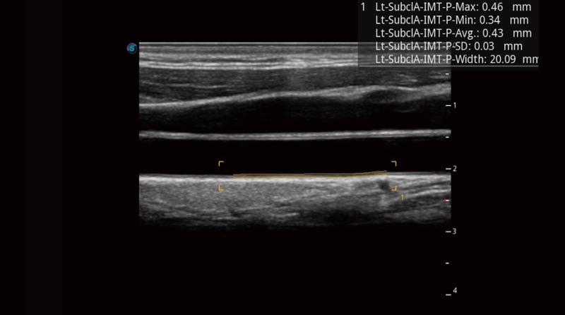
Auto IMT
Auto IMT makes the measurement of anterior and posterior intima-media thickness much easier with simple placement of the ROI.
-
![]()
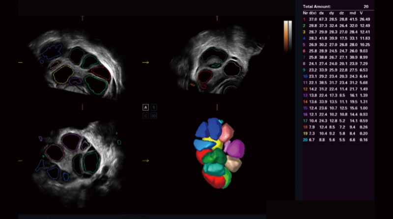
AVC Follicle
High efficiency of follicle analysis is achieved by AVC Follicle, a volume-data based automatic follicular calculation including the number and volume. Follicles are sorted by sizes in the results and rendered in different colors for better visualization.
Fast and Efficient Optimization
-
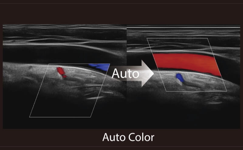
![]()
Auto B/C
Imaging parameter adjustment is now no more done in a laborious manner. Auto B/C helps to optimize the image quality under B and color Doppler mode within just one click. Multiple parameters such as gain, TGC, ROI position, steering angle, etc., are all included in this full automation method.
-
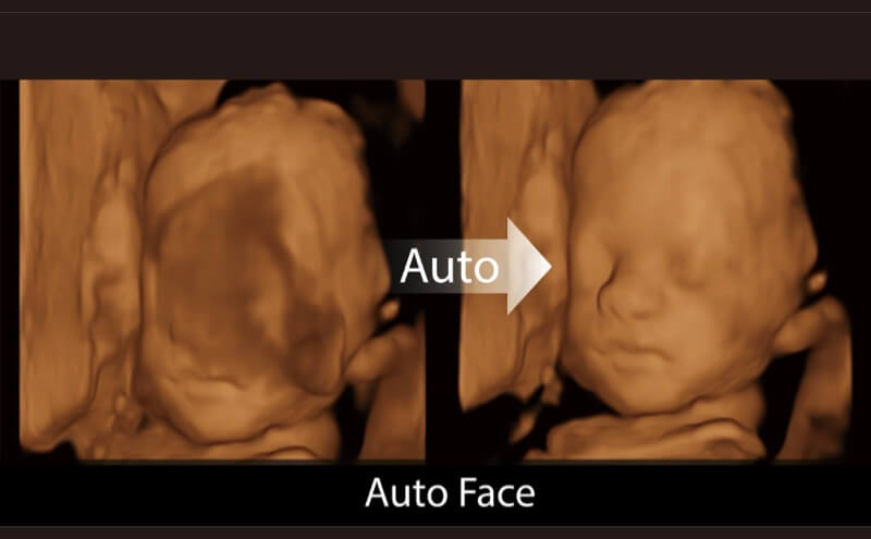
![]()
Auto Face
3D fetal face visualization is significant for face anomalies diagnosis. The removal of occlusions and artifacts, such as cord, placenta, uterus and extremities, can be simply accomplished by Auto Face to get an optimal view of fetal face.
Talented Features Inspire More Applications
Ultrasound is being versatile and taking on more and more clinical tasks. As a vanguard to help clinicians easily accomplish more, ELITE, is integrated with a comprehensive suite of advanced features covering General Imaging, OB/GYN, Cardiovascular and more.
MFI
MFI is an enhanced perfusion display enabled by the signal accumulation of contrast agents. It is useful for tracing small bubble populations, even in low-perfused and peripheral regions.
MFI Time
MFI Time provides a color coded parametric view, indicating the uptake time of contrast agents in different perfusion phases to better differentiate tissues.

S-Live Silhouette
Through the application of an artificial light source and shadowing effect, S-Live Silhouette sees through the surface and clearly delineates the outlines of bone, organs, cavities, vessel walls and other internal structures. It is a beneficial tool for identifying normal anatomy and diagnosing complex congenital malformations.

Color 3D
Available on color and power Doppler mode, Color 3D applies advanced rendering to blood flow to produce more intuitive and natural hemodynamics of vascular networks with speed and direction information, especially for umbilical cords.

Stress Echo
A straightforward template for clinicians to take multiple dynamic images at rest and after stress and make side by side comparison. Professional wall motion bulls-eye scoring and reporting is provided for further effective evaluation of cardiac muscle viability.

Myocardium Quantitative Analysis (MQA)
Precise quantitative measurement on myocardial mechanics is achieved by MQA based on real-time sensitive wall motion tracking. It provides global and regional assessment including strain, strain rate, displacement, velocity, etc.
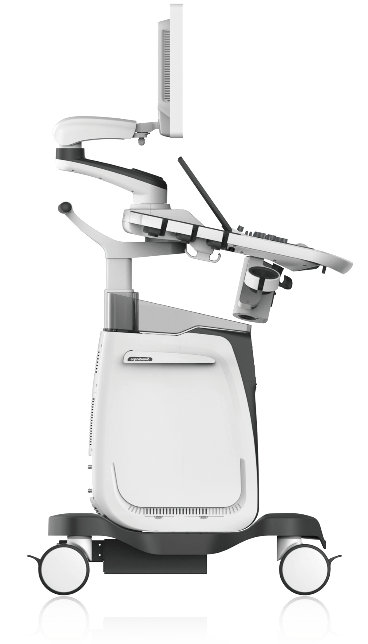
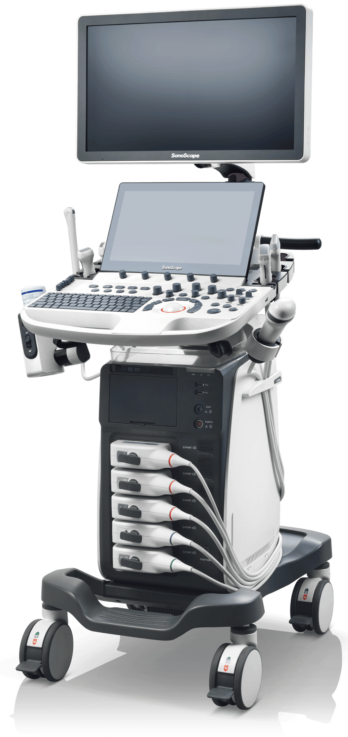
Easeful Experience within Easy Reach
Ultrasound is being versatile and taking on more and more clinical tasks. As a vanguard to help clinicians easily accomplish more, ELITE, is integrated with a comprehensive suite of advanced features covering General Imaging, OB/GYN, Cardiovascular and more.
-
![]()
Fully-articulating arm Easy adjustment for monitor position to enhance visibility.
-
![]()
High-resolution monitor & Touch screen 23.8 inch monitor (optional) and 13.3 inch touch screen for fatigueless view and smooth operation.
-
![]()
Intuitive user interface Straightforward layout effectively reduces keystrokes and manipulations. Customizablekeys increase flexibility for different user preference.
-
![]()
Flexible console Height-adjustable and rotatable console can basically satisfy any scanning requirements.
-
![]()
Compact build Slim and robust design offers enhanced mobility and easy accommodation even in difficult space.
-
![]()
Long-lasting capability Power management with a battery supporting 2 hours continuous scanning per charge in case of power failure.
Considerate User Interaction
-
Sono-Help
An inspiring tutorial displaying probe placement, anatomy illustration and standard ultrasound image examples. As a useful reference less experienced clinicians could rely on, Sono-help covers a variety of applications including liver, kidney, cardiac, breast, thyroid, obstetrics, vascular, etc.
-
Sono-Drop
Sono-drop provides a fast and convenient ultrasound image transmission between P40 ELITE and the patients’ smart devices. The bond between clinicians and patients are supposed to be strengthened through more frequent communication.
-
Sono-Synch
Real-time interface and camera sharing, enabled by Sono-synch, makes it possible to connect two ultrasound in a remote distance and perform remote medical consultation and tutorial.
Image Gallery
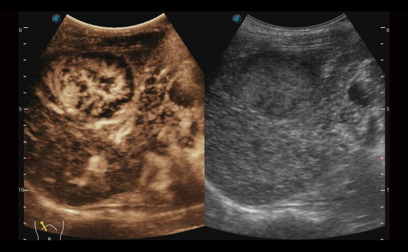
Fetal Abdominal with Single Crystal C1-6
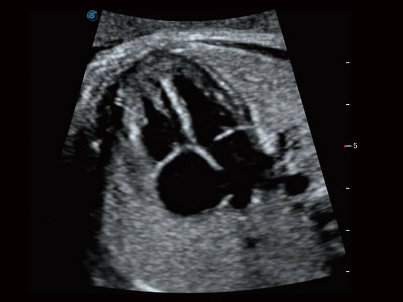
Fetal Heart with Single Crystal C1-6
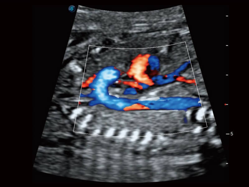
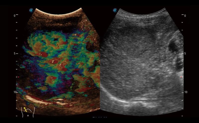
Fetal Circulatory System Blood Flow with SR-Flow

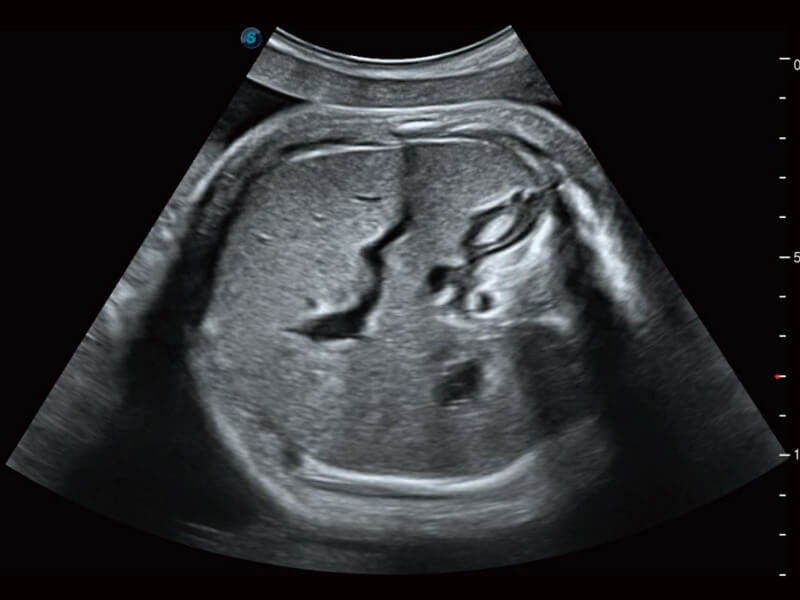
Lifelike Fetal Face with S-Live

Polycystic Ovarian Syndrome with 6V3

Hepatic Vessel with Color 3D

Thyroid Nodule with SR-Flow

Inferior Vena Cava with μ-Scan+

Tricuspid Regurgitation with Single Crystal S1-5